MEIOSIS
In unit 11 you learned that mitosis was
the division of a parent cell into two daughter cells which were identical to
each other and the parent cell. In this unit you will learn about meiosis. In
meiosis a parent cell will divide into daughter cells with half the number of
chromosomes as the parent cell. The only cells which use meiosis are the sex
cells, or gametes (sperm and egg). All other cells of your body go through
mitosis to divide. You will see that many of the stages of meiosis are similar
to mitosis, but, again, the important idea to remember about meiosis is that
the chromosome number will be cut in half in the daughter cells.
Sexual Reproduction
To reproduce sexually,
organisms need two cells to join. These two cells must have half the number of
chromosomes as all other cells of the body so that when they unite the new cell
will have a complete set of chromosomes for the new organism. These sexually
reproducing organisms have meiosis to provide a way of producing these cells
with half the number of chromosomes. Before you learn about the steps of
meiosis, you need to learn some of the terminology that is used with sexual
reproduction and meiosis.
“Cells”
The reproductive cells
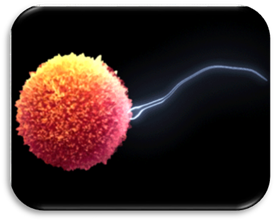
that meiosis produces are known as gametes.
The gametes are sperm and egg. When sperm and egg unite, a complete set of
chromosomes are formed in a process called fertilization.
The new cell formed as a result of fertilization is called a zygote. This zygote is the beginning of
the new individual. Because the zygote has traits of both parents (from the
sperm and egg), the new organism will not be exactly like either parent. Cells
that are specialized for producing gametes for sexual reproduction are known as
germ cells. All other cells of the
body are called somatic cells.
Somatic cells have nothing to do with sexual reproduction. If somatic cells
need to divide they will divide by way of mitosis.
Chromosome Number
All organisms have a
unique number of chromosomes. A dog has 78 chromosomes, a frog has 26, and
different species of ferns have over 500. Humans have 46 chromosomes. As you
can see with these examples, the chromosome number has nothing to do with the
complexity of the organism. However, the number of chromosomes is vital to the
organism. Too many or too few chromosomes and the organism may not develop
and/or function properly.
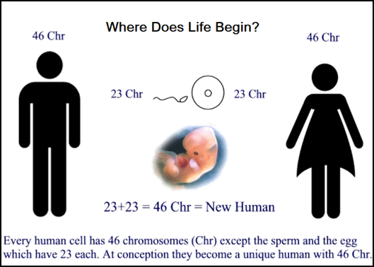
In terms of humans, we
have 46 chromosomes which are made up of 2 copies of 23 types of chromosomes.
One copy came from our father by way of the sperm and the other copy came from
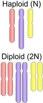
our mother by way of the egg. The cells (gametes) that have only one set of
each type of chromosome are called haploid.
The cells (somatic) that have two sets of each type of chromosome are called diploid. The symbol n is used to represent the number of
chromosomes in one set of chromosomes, or how many there are of each
type of chromosome. In humans, the haploid
number is n = 23, because the human
has 23 types of chromosomes. When you consider a somatic cell, it has 2 sets of
23 chromosomes so the diploid number
is 2n = 46.
“Chromosomes”
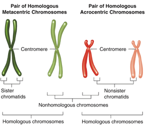
When looking at a diploid
cell you will find two of each type of chromosome. These two chromosomes of
each type are described as homologous chromosomes. Homologous chromosomes are similar in size and shape, and the genes
that they contain, however, the specific code of the gene will be different
between the two chromosomes. Even though eye color is controlled by more than
one gene we will use this as an example. On one of your mother’s chromosomes is
the gene for eye color. On your father’s homologous chromosome (same size,
shape, and genes as the mother’s) is also the gene for eye color. However, the
gene for eye color on the mother’s chromosome may be for blue and the gene for
eye color on the father’s may be for brown. They are homologous chromosomes
because they have the same size, shape, and genes, although they have different
forms of the gene.
When looking at the 46
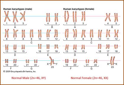
chromosomes of the human, there are two types of chromosomes. Autosomes are chromosomes with genes
that do not determine the sex or gender of an individual. Sex chromosomes are chromosomes with genes that do determine the
sex or gender of an individual. The sex chromosomes are referred to as “X” and
“Y” chromosomes. A male has the sex chromosomes “XY”, while a female has the
sex chromosomes “XX”. Keep in mind, humans are diploid organisms and have two
of each type of chromosome. So one “X” comes from the mother and the other “X”
or “Y” comes from the father.
Some scientists can
examine the chromosomes by looking at a karyotype. A karyotype is a picture of chromosomes. A human karyotype will show,
after it has been prepared and arranged, all homologous chromosome pairs lined
up from largest to smallest with the sex chromosomes placed last. Chromosome
pairs 1 to 22 are the autosomes and pair 23 are the sex chromosomes.
Stages of Meiosis
We will now look at the
stages of meiosis. This will explain how one diploid germ cell begins the
process and becomes four haploid gametes with half the number of chromosomes.
Meiosis involves two divisions of the nucleus called meiosis I and meiosis II.
In meiosis I the homologous chromosomes are separated producing two haploid
cells, but the sister chromatids are still attached. In meiosis II, the
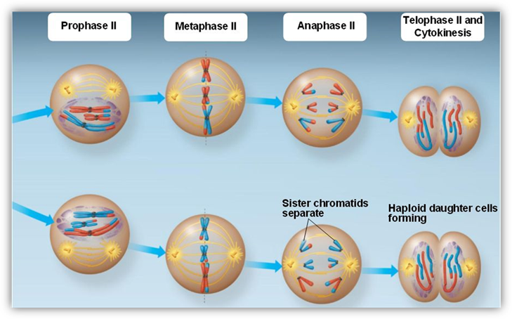
sister chromatids separate producing four haploid cells. Meiosis II is very
similar to mitosis as you will see. Meiosis I involves four stages (prophase I,
metaphase I, anaphase I, and telophase I), and meiosis II also involves four
steps (prophase II, metaphase II, anaphase II, and telophase II).
|
Stages
of Meiosis I |
|||
|
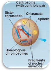
Prophase
I |
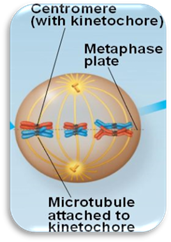
Metaphase
I |
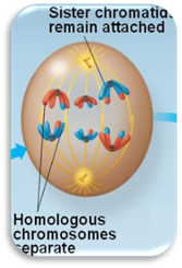
Anaphase
I |
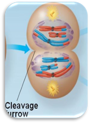
Telophase
I |
|
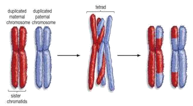
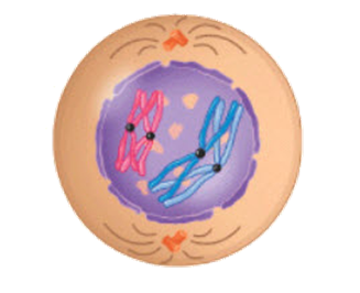
· Homologous
chromosomes form tetrads. · Crossing-over
occurs. |
· Tetrads
line up on the equator. |
· Tetrads
separate. |
· Cytokinesis
occurs. |
|
|
|
|
|
|
Prophase
I |
|
|
|
Metaphase
I |
|
In metaphase I, the tetrads line up on
the equator. It is not so much the centromeres lining up on the equator as it
is now the tetrad pair lining up on the equator. |
|
Anaphase
I |
|
In
anaphase I, the tetrads separate.
It is important to remember that the sister chromatids are still attached by
the centromere. This phase is just separating the tetrad. |
|
Telophase
I |
|
In
telophase I, the homologous
chromosomes, with their two chromatids still attached, are separated into two
new nuclei. The spindle breaks down. A new nuclear envelope reforms around
each set of chromosomes. In some cells the chromosomes will uncoil
(decondense). Cytokinesis also occurs during this phase to produce two new
cells. In the end of meiosis I, two cells are produced with half the number
of chromosomes, but the problem is the chromosome is still made of two
chromatids. Meiosis II is needed to split the centromere and separate the
chromatids. |
|
|
|
Stages
of Meiosis II |
|||
|
Prophase
II |
Metaphase
II |
Anaphase
II |
Telophase
II |
|
· New
spindle forms |
· Centromeres
line up on the equator. |
· Centromeres
separate. |
· Cytokinesis
occurs. |
|
|
|||
|
Prophase
II |
|
In
prophase II, the nuclear membrane
disappears, centrioles duplicate and move to opposite poles, and the spindle
forms. The DNA (chromosomes) are not copied between Meiosis I and Meiosis II,
as they were already copied before meiosis I.
[Remember there are two cells
going through meiosis II that came from the end of meiosis I]. |
|
Metaphase
II |
|
In
metaphase II, the centromeres line
up on the equator. |
|
Anaphase
II |
|
In
anaphase II, the centromeres split separating the chromatids to opposite
poles. Once the centromere splits the chromatids are now called chromosomes. |
|
Telophase
II |
|
In
telophase II, a new nuclear envelope forms around the chromosomes.
Cytokinesis also occurs during this phase to produce four new haploid cells (two new cells for each of the two cells
produced at the end of meiosis I). In the end of meiosis II, four cells
are produced with half the number of chromosomes from one diploid germ cell. |
UNIT VOCABULARY
REVIEW
Click on the Quizlet icon below to access the quizlet.com vocabulary flash
cards. Review the vocabulary before completing your assessment.
