CELLS
History of Studying Cells
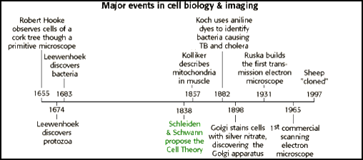
Because of the limitations of the human eye, much of the early
biological research concentrated on developing tools to help us see very small things.
As imaging technology became more sophisticated, biological discoveries
abounded. Below is a timeline detailing some of those major events in biology.
The Cell Theory
In 1838, Matthias Schleiden, a German
botanist, discovered and stated that plants are made of cells. In 1839, Theodor
Schwann, a German zoologist, discovered and stated that animals are made of
cells. In 1858, Rudolph Virchow, a German physician, stated that cells only
come from the division of previous existing cells.� This idea was important to recognize and
state because of the popular belief, at the time, of spontaneous generation.
Spontaneous generation dealt with the idea that living things could possibly come
from non-living things. If you recall, we mentioned in a previous lesson how
Louis Pasteur was the scientist that finally disproved spontaneous generation.
The �cell theory�, developed by Schleiden, Schwann, and Virchow, changed
biology and cell biology research forever.
The
cell theory states that:
�
Living
things (organisms) are made of one or more cells.
�
Cells
are the basic unit of structure and function in organisms.
�
All
cells arise from existing cells.
The cell theory also provides us with an operational definition of "life" as it is one of the characteristics of life mentioned in a previous lesson.
Cell
In biology, the basic unit of which all living things are
composed is called a cell. As the smallest units retaining the fundamental
properties of life, cells are the �atoms� of the living world. A single cell is
often a complete organism in itself, such as a bacterium or yeast. Other cells,
by differentiating in order to acquire specialized functions and cooperating
with other specialized cells, become the building blocks of large multicellular
organisms as complex as the human being. Although they are much larger than
atoms, these building blocks are still very small. The smallest known cells are
a group of tiny bacteria called mycoplasmas;
some of these single-celled organisms are spheres about 0.3 micrometers in
diameter, with a total mass of 10-14 gram�equal to that of
8,000,000,000 hydrogen atoms. Human cells typically have a mass 400,000 times
larger, but even they are only about 20 micrometers across. It would require a
sheet of about 10,000 human cells to cover the head of a pin, and each human
being is composed of more than 75,000,000,000,000 cells.
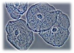
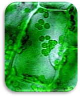
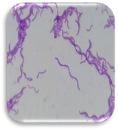
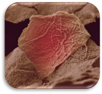
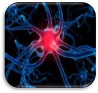
Cell Size and Shape
|
|
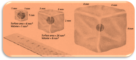
Not all cells need to stay small, some cells can survive larger
when compared to others.� Larger cells
need to have shapes that increase the surface area so that enough exchange in and
out of the cell can occur. A cell can only grow large in one dimension and
still survive.� Some large cells are long
and skinny or broad and flat. We tend to always see models of cells, in
pictures and hand-held models, as circular objects, when in reality all cells
differ in size and shape. Below are some pictures of different types of real
living cells and how they differ in shape.
|
|
|
|
|
Cell Structure
All cells, regardless of type, have the same five features in
common. Those five features are cell
membrane, cytoplasm, cytoskeleton, ribosomes, and DNA.
Cell
Membrane is the cell�s outer boundary that acts as a barrier between the
outside and inside of a cell. We will discuss more about the cell membrane�s
structure and function in a future unit.
The cytoplasm is the
material from the nucleus to the cell membrane or, if no nucleus is present,
everything within the cell membrane solid and liquid.� The cytoplasm consists of the fluid, which is
known as cytosol, with all of the dissolved particles in it, and all of the
structures suspended in it. Cytoplasm contains molecules, ions, water, and
every cell organelle, except the nucleus. The liquid of the cytoplasm acts like
a buffer, maintain a pH that promotes life, helps chemical reactions to work optimally.
The cytoskeleton is
what enables the cell to have a particular shape, organize the parts within,
and move either as a whole cell or the parts within. The cytoskeleton is
primarily mentioned as a feature of a cell which has a nucleus (eukaryote), but
similar structures have also been found in cells without a nucleus
(prokaryotes). When one looks at one single-cell organism, the ameba, you see a
blob that can alter its shape to suite its needs of movement, eating, and
disposing of waste. But many cells have specific shapes. Cytoskeletons are networks of proteins
the helps the cell maintain its shape without the need of extra energy to
contract the cell membrane. The cytoskeleton
is composed microtubules, microfilaments, and intermediate fibers.
Microtubules are hollow, protein
tubes that act as "tracks" along which organelles can move through a
cell. Microfilaments are long,
thin protein fibers that help cells move, change shape, and/or provide some
shape and structure to the cell. Microfilaments
are much thinner than microtubules,
7nm and 25nm respectively. Intermediate fibers are moderately thick (size is
between microtubules and microfilaments) and mainly anchor organelles and
enzymes to certain regions of the cell.
All cells have ribosomes
which are responsible for producing the proteins of a cell. Ribosomes may have one of the most
important jobs within cells. Ribosomes
carries out the job of assembling proteins, based upon the blueprint found in
the DNA molecule, by linking together amino acids. Ribosome uses both mRNA,
and tRNA to make proteins. Ribosomes are found on sections of an
organelle called the endoplasmic
reticulum or floating independently in the cytosol. When the ribosome make proteins, the proteins
directly enter the endoplasmic reticulum
(if present).
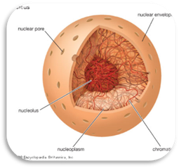
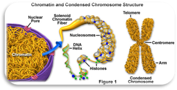
Lastly, all cells have DNA
(deoxyribonucleic acid) which is the genetic material responsible for
providing the instructions for making proteins, regulating cellular activities,
and enabling the cell to reproduce.
![]() ��� The Characteristics of Cells (01:21)
��� The Characteristics of Cells (01:21)
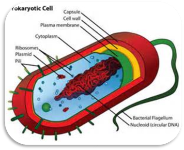
Prokaryote and Eukaryote Cells
There are two basic types of cells that exist, prokaryote and eukaryote. A prokaryote is a cell which does not
have a nucleus or membrane-bound organelles.� A eukaryote
is a cell which has a nucleus and membrane-bound organelles. The only
prokaryotes that exist are the single-celled (unicellular) organisms known as
bacteria. All other organisms, whether it be a protist, fungus, plant, or
animal and unicellular or multicellular, are eukaryotes.
|
|
|
|
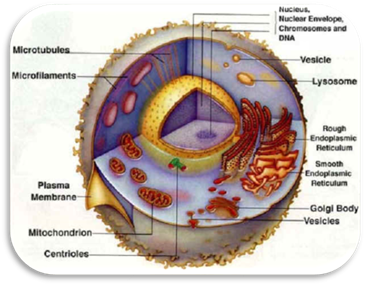
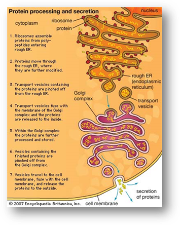
Endoplasmic Reticulum
|
The endoplasmic reticulum is a long internal system of
continuous membranes, running throughout a cell and usually attached to the
nuclear envelope. The section of the endoplasmic
reticulum that has ribosomes is
called the rough endoplasmic
reticulum (rough ER).
It is called rough because of the rough, bumpy look that the ribosomes give
the surface of the endoplasmic reticulum. Where you do not have ribosomes, the endoplasmic reticulum is called the smooth endoplasmic reticulum
(smooth ER). Within this section of the endoplasmic reticulum, specialized enzymes can be found, making
things like lipids and breaking down toxins. The polypeptides (beginning of a
protein) that are made in a ribosome
found on the rough endoplasmic
reticulum are then transported to the end of the rough ER membranous channel
and pinched off by the ER membrane into a vesicle (transport
vesicle). The vesicle transports the newly made substance just as an envelope
transports a letter through the mail. These vesicle-enclosed polypeptides are
then transported to the Golgi
apparatus. Golgi Apparatus The Golgi apparatus
(complex) finishes the proteins, by modifying the polypeptide (adding
carbohydrates and lipids to the protein). From the Golgi apparatus, proteins are finally sent on their way through
the endoplasmic reticulum, to
the outside of the cell by way of another vesicle which again is just the
membrane of the now Golgi apparatus pinched off. This process of making a polypeptide by way of the Ribosome to the
Endoplasmic Reticulum to the Golgi Apparatus is sometimes referred to as
�protein processing� and/or �protein transport�. Vacuoles An advantage of eukaryotes over prokaryotes
is the membrane bound organelles. This is particularly the case when the cell
makes too many proteins, like digestive enzymes. Vacuoles are the storage organelles of the cell. Vacuoles hold water, salts,
proteins, carbohydrates, and sugars. Specialized vacuoles may contain very strong digestive enzymes. In this
case, we call the organelle lysosomes.
Lysosomes contain powerful enzymes the can rapidly breakdown proteins,
nucleic acids, lipids, and carbohydrates, so the lysosome acts as the stomach of the cell. By breaking down these
biomolecules, the remaining materials can now be recycled by the cell. Those
organelles that hold enzymes that can convert fats into carbohydrates, or can
convert hydrogen peroxide into water are called peroxisomes. Some
protists have specialized vacuoles known as contractile vacuoles which pump excess water out of their cell.
By pumping out excess water, these cells can now balance the concentration of
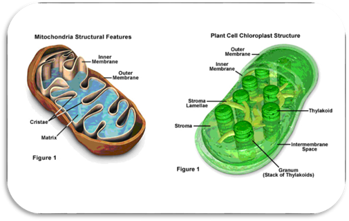
salt and other molecules in an effort to maintain homeostasis. Energy Producing
Organelles Two important organelles involved in converting
energy into a usable form are chloroplast
and mitochondrion. Chloroplasts
contain molecules and carry out a process (photosynthesis) that converts the
energy from sunlight into sugars. Mitochondria (nicknamed the
�powerhouse� of the cell) contain enzymes that can convert the energy in
sugars and other high-energy molecules to ATP (the cells fuel) which is the cell�s only usable form of
chemical energy. Once the energy stored in sugar has been converted to ATP, the cell has the extra energy
that it needs to run complex processes. More details about the reactions and
processes that take place in the mitochondrion and chloroplast will be
discussed later.
|
|
The following video
profiles the structure of a eukaryotic cell, describing the form and function
of each part of a cell. The program covers the phospholipid bilayer,
cytoskeleton, cytoplasm, mitochondria, lysosome, nucleus, and the rough and
smooth endoplasmic reticulums.
|
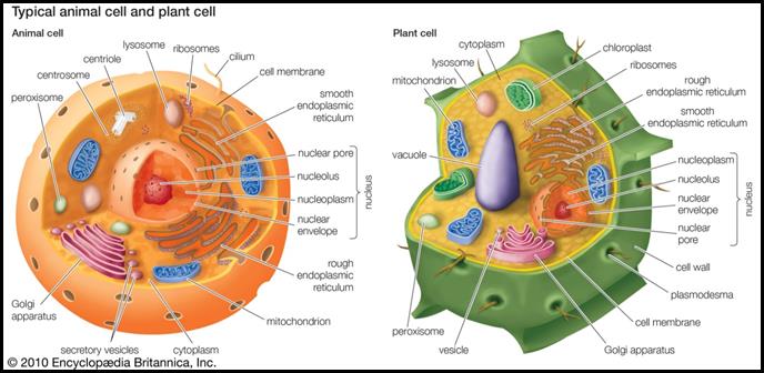
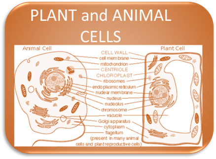
Animal
Cell vs Plant Cell
Plant cells have three structures that animal
cells do not. Plant cells have a central vacuole, chloroplasts, and cell
wall.� We have already discussed
vacuoles.� A central vacuole is the same
as a vacuole, however, it is much larger in a plant cell as it may take up as
much as 90% of the volume of the plant cell�s center. We also already mentioned
chloroplasts as providing a means of chemical energy.� The cell wall in a plant cell is located
outside of the cell membrane and is only found in a plant cell and not an
animal cell.� The plant�s cell wall
provides structure, support, and protection for the plant cell. The primary
component for the plant�s cell wall is a complex carbohydrate named cellulose.
Additional
practice: Print out the following worksheets to see if you can correctly label
all the parts of the cell. This is practice only!
Printable Animal Cell Worksheet
Printable Plant Cell Worksheet
Plant and Animal Cell Worksheet
UNIT VOCABULARY
REVIEW
Click on the Quizlet
icon below to access the quizlet.com vocabulary flash cards. Review the
vocabulary before completing your assessment.

 ��� Now answer questions 1
through 25.
��� Now answer questions 1
through 25.