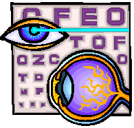
THE HUMAN EYE
Unit Overview
This unit will help you better understand what your eye is made of and exactly
how it works. You will also learn common eye problems and some things you can
do to prevent these problems. Good luck.
How the Eye Works
Vision is a complex
sense composed of many elements. The human eye, elegant in its detail and
design, represents a gateway to the process we call vision. The eyeball, or
globe, is spherical in shape and about 1 inch across. It houses many structures
that work together to facilitate sight.
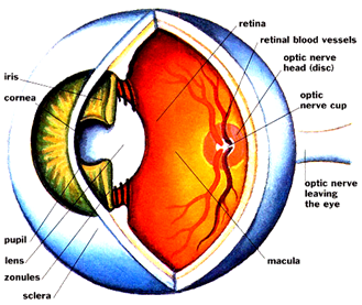
The human eye is
comprised of layers and internal structures, each of which performs distinct
functions. The outside layer of the eye is comprised largely of a tough, white,
protective tissue called the sclera. The sclera helps maintain the shape of the
eyeball. At the front of the eye is an equally tough but clear structure called
the cornea, which is responsible for letting light into the eye and bending light.
Going from outside to
inside, the next layer of the eye is the choroid, which carries the blood
supply necessary to nourish the eye's internal structures. Finally, there is
the layer called the retina, lining the inside of the eye, which is sensitive
to light and receives stimulation to its specialized cells.
The eye has a number
of protective features. The eyelids, eyelashes and eyebrows are all designed to
protect the eye from dirt and dust that might enter it and cause damage. The
globe sits inside the orbital cavity, a bony pocket lined with fatty tissue as
a cushion. Together these provide additional protection against injury. Six
muscles attach at various points to the sclera and enable the globe to move in
many directions inside the orbit.
In order for vision
to take place, a succession of processes must occur involving the structures
within the eye and the brain:
The first part of
this chain is that light rays must travel through the eye to ultimately focus
on the retina. There are a number of structures involved in the bending or
refracting of light so that it focuses properly. Light first passes through the
clear cornea at the front of the eye, and then through a watery substance
called the aqueous humor which fills the small chambers located behind the
cornea. As light continues on its pathway it passes through the pupil, a round
opening in the center of the iris. The iris is the part of the eye that gives
the eye its color. It also is made up of specialized muscles that are able to
change the size of the pupil from very small (about 2 mm) to large (about 8
mm), regulating the light that is entering.
The next structure
light will penetrate the lens, another clear, layered structure shaped like a
large lentil (about 10 mm in diameter) that is attached to muscles which
contract or relax to change the shape of the lens. The changing lens shape
helps light to be focused in response to the need for clarity. (The loss of
this focusing ability as humans age – a natural occurrence – is the reason that
many adults over 40 years old need reading glasses.) Once through the pupil and
lens, the light then passes through the larger posterior (back) portion of the
eye that is filled with a clear, jelly-like substance called the vitreous
humor. From there, the light will come to the retina, where the rod cells and
cone cells will be stimulated to set off a chain of split-second chemical
reactions converting light to electrical impulses. The cone cells (about 7
million in number) are located in greatest concentration in the small, central
part of the retina called the macula. This area is responsible for producing
sharp, detail vision and color vision. The rod cells (numbering about 100
million) are found in the peripheral retina, away from the macula. These cells
provide vision in dim light.
Even if all of the
structures of the eye work perfectly, what we know as vision cannot happen
without the brain's interpretation of the electrical impulses sent by the
retina. The optic nerve is the bundle of retinal fibers that exits the back of
the eye and transports electrical impulses to the brain where they are
interpreted in the primary visual cortex.
When all parts of the
visual system are working, the eyes can move together, can adapt to light and
dark, perceive color and accurately evaluate an object's location in space.
They are sensitive to differences in contrast, and can also provide detail
vision, which is measured as visual acuity. By convention, we know
"normal" visual acuity to be reported as 20/20. As the bottom number
of this expression gets higher, it tells us that the vision is poorer than
"normal." For example, the start of the range known as "legal
blindness" is represented by the visual acuity finding of 20/200. One way
to understand the meaning of this finding is that the eye being tested sees at
20 feet what the "normal" eye would see at 200 feet. People whose
vision is in the category of "legal blindness" may still be able to
use vision to do some of the things they need to do.
All eyes are not the
same, nor are they all perfect. Some eyeballs are too long or have too much
focusing power, causing the person to be myopic (nearsighted). Others are too
short or have too little focusing power, and the result is hyperopia
(farsightedness). Some eyeballs may have uneven curvature, called astigmatism.
Options for correcting these "mechanical" problems are standard
eyeglasses, contact lenses or refractive surgery. Other problems may be caused
by disease or injury, and are not correctable by conventional means. People
whose vision is irreversibly impaired due to diseases such as macular
degeneration, glaucoma, cataract, diabetic retinopathy and others can be helped
by vision rehabilitation.
![]() Properties of Vision: How the Eye Senses
Information (04:19)
Properties of Vision: How the Eye Senses
Information (04:19)
![]() Now answer questions 1-7.
Now answer questions 1-7.
Common Eye Problems
Myopia
(Nearsightedness) is a refractive error where distant objects come into focus
in front of the retina of the eye as a result of longer than normal eye-ball,
so the distant object appears blurred to the myopic eye. A bent concave lens is
used to bring the focus on the retina to restore clear distance vision.
However, the myopic
eye can still see near objects clearly without any correction depending on the
distance of the near objects in relation to the degree of myopia.
Hypermetropia (Long-sightedness)
Hypermetropia (Long-sightedness) is a refractive
error where distant object comes into focus behind the retina when the eyes are
relaxed. In adults, this condition will cause deterioration of distance vision
and near visual problem due to the gradual loss of focusing ability of the eye.
A bent convex lens is used to bring the focus back onto the retina. In
children, distance vision is often not affected because of the active and
strong focusing mechanism, the eyes have ability to
bring the focus back onto the retina. However, for higher degrees of hypermetropia, it may cause blurred vision, headache, and
even converging squints. These problems will be aggravated when doing near
works which required greater focusing power. It's important to detect and
correct the medium to high degrees of hypermetropia
for children under 6 years old. If uncorrected, it can lead to Amblyopia. (lazy eye).
Click on the
following link to learn more about Lazy Eye in Children. http://www.childrensvision.com/lazyeye.htm
Astigmatism
Astigmatism is a
refractive error where distant object comes into two focal lines perpendicular
to one another. These lines may lie in any orientation and any position in
relation to the retina. This is primarily due to the aspherical
(toroidal) corneal shape. A bent lens with a toric surface is used to correct this condition.
Astigmatism may occur
by itself or may occur with one of the refractive errors but in most cases are
less than 2.50 dioptres. Although for a similar
degree of defect, astigmatic eyes suffer less blurriness than the other
refractive errors, uncorrected astigmatism often leads to eyestrain and headache
due to the focal lines formed in the eye cause the eye's focusing mechanism
under strain as it constantly exert effort for best possible focus.
Uncorrected
astigmatism on young children can also lead to Amblyopia.
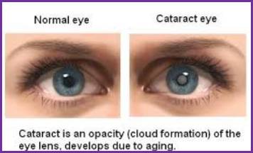
Cataracts
A cataract is a clouding
in the eyes natural lens, which is normally crystal clear. It is not a tumor or
growth over the eye. Most cataracts progress and eventually hamper vision. They
develop as part of the aging process and are not usually a problem until the
60s or 70s. Studies have shown that prolonged exposure to sunlight can play a
role in hastening cataract development. Everyone will get cataracts if they
live long enough. If you have a cataract, you might notice blurring or dimming
of vision, or a halo or haze around lights, especially at night. You may not
notice even an advanced cataract if your other eye sees well. Eye pain,
headaches, or eye irritation are not symptoms of
cataracts.
The only treatment
for cataract is removal of the cloudy lens, replacing it with a new synthetic
"intraocular lens implant". Cataract surgery is one of the safest and
most effective operations today. The most modern "Clear Corneal Cataract
Surgery" is performed by Dr. Craig as same day surgery at Henderson
Memorial Hospital, Laird Memorial Hospital in Kilgore and East Texas Medical
Center in Carthage. The surgery is performed under local or eye drop
anesthesia. This state of the art surgery is 90-95% successful.
![]() Eye Defects (02:04)
Eye Defects (02:04)
Glaucoma
Glaucoma is the term
given to the set of eye diseases in which the intraocular pressure within the
eyeball may be increased and progressive damage to the optic nerve results. The
optic nerve is like a main telephone trunk, transmitting images from the retina
of the eye to the brain. As glaucoma slowly damages the optic nerve, blind
areas develop that can progress to loss of vision or even total blindness,
usually painlessly. That is why we call it "The Sneak Thief of
Sight".
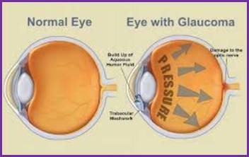
Ninety percent of
patients with glaucoma have the open angle or chronic type. It usually affects
both eyes, often one before the other and in mid or late life. It may be
hereditary, is not contagious, and is not related to cancer. High pressure in
the eyes is not the same as high blood pressure. As part of your routine eye
exam, Drs. Craig and Pinkerton will do a painless test called tonometry to
measure your eye pressure. Elevated pressure is one sign of glaucoma, but other
tests to confirm this diagnosis may be needed. A visual field will measure your
side vision and examination of the retina and optic nerves is an integral part
of your exam. Appointments with Drs. Craig or Pinkerton every few months will
follow, so that they can monitor your condition, in order to prevent
progression of the disease. Eye drops to lower pressure can be prescribed and,
in some cases, laser surgery or filtering surgery may be required.
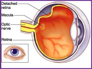
Detached Retina
Usually the retina is
attached to the inner surface of the eye. if there is
a tear or hole in the retina then fluid can get underneath. This weakens the
attachment so that the retina becomes detached - rather like wallpaper peeling
off a damp wall. When this happens the retina cannot compose a clear picture
from the incoming rays and your vision becomes blurred and dim.
![]() Now answer questions 8-12.
Now answer questions 8-12.
Eye care
The following
suggestions are for information purposes only and meant for healthy and normal
eyes which do not have any complications. Those with specific eye related
problems should consult their ophthalmologist or health care providers before
doing any particular exercise.
1. Adequate rest and
relaxation are very important for the proper care of your eyes. Stress and
strain result in building up of pressure on the optic nerve, the eye muscles
and the retina as well as cause changes in the flow of blood in the veins that
supply blood to the eyes. So keep yourself and your eyes well relaxed most of
the times. If you feel tired and stressed take rest, so that you will feel
charged and ready to face the world again.
2. During the day,
especially when you have no work to do, give rest to your eyes intermittently.
You can do this even amidst busy schedule taking a moment off. The best way to
rest your eyes is to temporarily close them. Do this whenever possible for a
few minutes each time. By doing this you will be helping your eyes to work more
efficiently and providing the nerve cells the much need temporary relief.
(Well, you won't practice this when you are driving a car or walking down a
road or climbing up or down a stair case, etc.!)
3. Do not practice
self-medication when there is any problem in your eyes. Consult always a
qualified ophthalmologist. Do not use any eye drops or ointments or medicines,
unless prescribed and recommended by your ophthalmologist. This may not save
your purse and your time, but definitely your eyes!
4. We do not like
people staring at us, do we ? (Unless of course by a
person whom you are attracted to!) Blink your eyes frequently and help your
eyes relax and remain moistened. People with increased eye sight have a
tendency not to blink for longer durations. See whether you too have developed
this habit unconsciously.
5. Do some eye
exercises if possible every day. There are many and your ophthalmologist can
suggest you some specific ones. One good exercise I found useful is the palming
exercise. In this exercise you sit comfortably, close both your eyes, cover
your eyes lightly (no pressure whatsoever) with your palms and keep looking
into the darkness in front of your eyes, in a complete state of relaxation,
without forcing your mind to think any particular line of thoughts or
subjecting yourselves to any strain. If you practice this exercise sincerely
you will soon see the results for yourself.
6. If you have vision
related problems, and if you are a middle aged person, keep your eyes examined
by a qualified ophthalmologist at least once in a year.
7. Avoid looking at
the sun directly even for a moment at any time of the day. The glaring light of
the sun is said to be very harmful to your eyes, especially to the cells in
your retina. It is also advisable to use sunglasses whenever you feel that the
light is causing irritation in your eyes.8. But it is useful to close your eyes
completely and turn your face towards the sun. The warmth of the sun light will
increase the flow of blood in your eyes and stimulate the nerve cells in your
eyes. When you do this please see that no light enters your eyes directly.
8. You must have read
this a hundred times and even got tired of it. But I must state it here again.
Eat plenty of vegetables and food items that contain Vitamin A
: eggs, fish, milk, leafy vegetables, carrot, to suggest a few.
9. Keep sharp
objects, harmful toys (like toy pistols that throw objects, dart boards, arrows
and bows etc.) and similar devices away from your children, if you want to
protect their eyes as well as yours. They have a tendency to play with them
with great enthusiasm, without bothering about the consequences!
10. Regular exercise,
moderation in your habits, positive state of mind, loving and caring nature,
contentment in life, cleanliness, harmony in your environment and in your
actions, a certain degree of interest in leading a meaningful and useful life,
etc., also contribute greatly to your overall health and thereby to the general
welfare of your eyes also.
![]() Now answer questions 13-15.
Now answer questions 13-15.

Below are additional educational resources and activities for this unit.
Unit 7 The Human Eye Puzzle Key