THE RESPIRATORY SYSTEM
In this unit you will trace the
path of the vital process of taking in oxygen and excreting carbon
dioxide. You will detail the organs as
well as the parts of the organs involved in this process. Good luck!
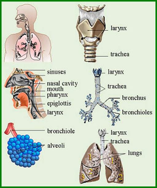
THE HUMAN RESPIRATORY SYSTEM
Take a
nice deep breath. Inhale through the nose, exhale through your mouth.
Aaaaahh........... Inhale, exhale. Inhale, exhale.
Inhale, exhale.
Hey
--- wake up ! Time to learn about the system of organs
that's responsible for taking in oxygen and getting rid of carbon dioxide ...
the respiratory system.
First, allow me to list the structures
and organs that together make up the respiratory system. Your job is to write
them down in the order that air passes through them as it is inhaled.
|
1.
alveoli 2.
bronchiole 3.
bronchus 4. larynx 5. lung 6.
pharynx 7. nasal
cavity 8.
nostril 9.trachea 10.
diaphragm |
Now let's take a look at what
those respiratory structures look like.
![]() Functions of the Respiratory System
Functions of the Respiratory System
You are now to identify and label those
same organs and structures that we listed just a minute ago?
|
alveoli (air sacs) |
bronchioles |
bronchus |
diaphragm |
|
larynx |
lung |
pharynx |
nasal cavity |
|
nostril |
trachea |
|
|
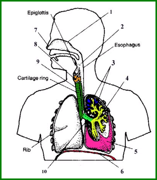
Click on the PDF to see answers:
Notes:
·
The esophagus is part
of the digestive system. It is the tube leading to the stomach.
·
The epiglottis is a
flab of flesh (tissue) that covers the trachea (windpipe) when you swallow so
that food doesn't "go down the wrong pipe". in
other words, the epiglottis prevents choking.
·
The lungs lie (well
protected) behind the ribs in the chest cavity.
·
a membrane called the pleura surrounds each lung
Here is a summary of the functions of each Respiratory Structure
|
STRUCTURE |
FUNCTION |
|
nose / nasal cavity |
warms,
moistens, and filters air as it is inhaled |
|
pharynx (throat) |
passageway
for air, leads to trachea |
|
larynx |
the
voice box, where vocal chords are located |
|
trachea (windpipe) |
tube
from pharynx to bronchi |
|
bronchi |
two
branches at the end of the trachea, each lead to a lung |
|
bronchioles |
a
network of smaller branches leading from the bronchi into the lung tissue and
ultimately to air sacs |
|
alveoli |
the
functional respiratory units in the lung where gases (oxygen and carbon
dioxide) are exchanged (enter and exit the blood stream) |
|
diaphragm |
This
muscular structure acts as a floor to the chest (thoracic) cavity as well as
a roof to the abdomen. It helps to expand and contract the lungs, forcing air
into an out of them. |
|
lung |
The
lungs are the essential organs of respiration. The main function of the lungs
is to exchange carbon dioxide for oxygen and vice versa. Each lung is enclosed separately within two
membranes, like a balloon inside a bag inside. |
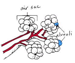 Let's take a closer look
at the air sacs (alveoli) in the lungs.
As the bronchioles branch out into
smaller and smaller and smaller and smaller and smaller tubes, they eventually
lead to microscopic clusters of alveoli, which are referred to as air sacs. You
can think of the air sacs as a bunch of grapes, with each individual grape
representing a single alveolus like this:
A close-up of the air sacs, which
are located at the ends of the bronchioles. Each "air sac" is
comprised of a cluster of alveoli. The red structures represent blood vessels
leading to and from the air sacs.
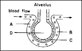
This is an even closer look at an
alveolus. Notice that the wall of an alveolus is only one cell thick. This
allows gases to diffuse into and out of the alveoli.
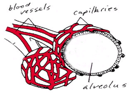 Also notice that the alveoli are surrounded by capillaries
so that oxygen and carbon dioxide can be exchanged between the lungs and the
blood. Oxygen in the alveolus can diffuse into the bloodstream (and be
transported throughout the body) and carbon dioxide in the bloodstream can
enter the alveoli (and then be exhaled).
And now a few words about
breathing!
There are no muscles in your
lungs. They do not actively pump air in unit out, in unit out. The muscle
responsible for breathing actually lies below the lungs. It is like a rubber
sheet that separates your chest cavity unit your abdominal cavity. It's name is diaphragm. When you
inhale, the diaphragm contracts unit moves downward, which
creates more space in your chest cavity unit draws air into the lungs. When you
exhale, the diaphragm relaxes unit moves upward, forcing
air out of the lungs. A common demonstration of the mechanics behind breathing
involves a bell jar, some glass tubing, and a couple of balloons. Like so:
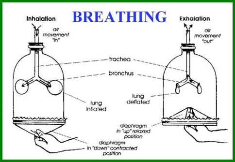
If you look closely at the right
side of the diagram, which represent exhaling, you can see how the Heimlich
maneuver works. The Heimlich maneuver is the "bear hug" that helps to
dislodge food from the windpipe of a choking victim. By pushing upwards below
the victims ribs, the diaphragm is forced up and air is forced out of the
trachea, hopefully with enough "UmmppHH" to
remove the blockage. See, biology can save your life.
One more
tidbit: hiccups are muscle spasms in the diaphragm.
|
Malfunctions and Diseases of the Respiratory
System |
|
|
asthma |
severe
allergic reaction characterized by the constriction of bronchioles |
|
bronchitis |
inflammation
of the lining of the bronchioles |
|
emphysema |
condition
in which the alveoli deteriorate, causing the lungs to lose their elasticity |
|
pneumonia |
condition
in which the alveoli become filled with fluid, preventing the exchange of
gases |
|
lung cancer |
irregular
and uncontrolled growth of tumors in the lung tissue |
Keep
your lungs happy and healthy --- keep them SMOKE FREE.
To learn more about the Respiratory System click on the PDF link and print out
the worksheet.
Respiratory
Worksheet PDF File
![j0371082[1]](HEALTHU06The_Respiratory_System_image017.png) Answer questions 1-20
Answer questions 1-20

Below are additional educational resources and activities for this unit.
Unit 6 Respiratory System Graph
Unit 6 Respiratory System Matching
Unit 6 Respiratory System Matching Key