MITOSIS
 Through
our work with previous lessons, you have now learned about the different types
of cells, the parts of a cell, the membranes of a cell, how substances move in and
out of a cell, and some of the most important metabolic processes in cells. In
this unit you will learn how a cell divides or reproduces. There are two ways
that cells, or more specifically a nucleus, can divide or reproduce: mitosis or
meiosis. This unit will examine mitosis while the next unit will look into
meiosis. As the human body grows larger, the cells do not necessarily become
larger. Instead, the cells become more numerous. Of the over 25 trillion
cells that make up the adult human body, about two trillion replacement cells
are produced every day. The new body cells are exact copies of the cells they
replace.
Through
our work with previous lessons, you have now learned about the different types
of cells, the parts of a cell, the membranes of a cell, how substances move in and
out of a cell, and some of the most important metabolic processes in cells. In
this unit you will learn how a cell divides or reproduces. There are two ways
that cells, or more specifically a nucleus, can divide or reproduce: mitosis or
meiosis. This unit will examine mitosis while the next unit will look into
meiosis. As the human body grows larger, the cells do not necessarily become
larger. Instead, the cells become more numerous. Of the over 25 trillion
cells that make up the adult human body, about two trillion replacement cells
are produced every day. The new body cells are exact copies of the cells they
replace.
New cells are small in
size and therefore have a higher surface area-to-volume ratio. As the cell
grows it produces more material and substances inside of itself making it
larger. As the cell grows larger and the surface area-to-volume ratio becomes
smaller the cell must divide or reproduce to survive.
The need for cell
division of new cells is not only necessary for growth but also for replacement
and repair. For example, the skin cells of the human body are constantly lost
from the outermost layer and need to be replaced. In addition, when you have a
cut in the skin of your finger, you need new cells to repair or heal the
damaged skin.
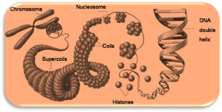
Chromosomes

Before we look at
mitosis, you need to understand the different ways DNA can be organized.
Remember that DNA controls all of
the activities within a cell and, in eukaryotes, DNA is found in the nucleus. DNA is divided into units of
differing lengths to code for RNA which in turn will code for a protein (to be
discussed in detail later). These units of DNA are called genes. DNA is made of thousands of genes. In order for this long
DNA molecule to fit inside the nucleus it must be condensed as much as
possible. The DNA coils and is packaged into a structure called a chromosome.
|
PROKARYOTIC
CHROMOSOME |
|
Recall that
prokaryotes do not have a nucleus. The DNA of prokaryotes is found in the
form of a circular loop floating in the cytoplasm. This chromosome consists
of DNA that has been twisted several times within its loop. |
|
EUKARYOTIC
CHROMOSOME |
|
|
|
As was already
mentioned, the DNA of a eukaryote is packed in the nucleus. Eukaryotic cells
contain more genes than prokaryotic cells so the DNA needs to be greatly
condensed. When the DNA and proteins associated with it are found scattered
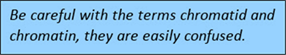
throughout the nucleus it is called chromatin.
Even though the DNA is found scattered it is still coiled or condensed to
allow all of it to fit within the nucleus. This coiling involves proteins
called histones. Eight histones create
a center core for the DNA to wrap or coil around. There will be many histones
in succession for the DNA to wrap around. The term nucleosome is used for one set of histones |
|
|
|
PROKARYOTIC
CELL DIVISION |
|
|
|
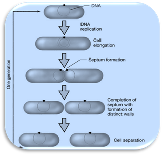
Prokaryotes,
having no organelles or nucleus to deal with, makes prokaryotic cell division
simpler than eukaryotic cell division. The circular DNA of the prokaryote will
attach to the inner cell membrane. Once attached, the DNA loop will be copied
with the copied loop also being attached to the inner cell membrane. The cell
membrane then begins to divide or pinch in between the two DNA attachment
points. The cell will continue to grow to provide enough material for the two
daughter cells. The cell wall will also form around the membrane that is
dividing the cell in half between the DNA attachment points. When the cell
membrane and cell wall finish separating the cell in half, two new
prokaryotic cells have now been produced each with its own DNA. This process
of cell division in a prokaryote is known as Binary Fission.
|
|
EUKARYOTIC CELL
DIVISION |
|
Eukaryotic
cell division is more complicated than prokaryotic cell division. In
eukaryotic cell division, enough organelles must be available for the two daughter
cells, along with having a nucleus containing an identical copy of DNA. |
Cell
Cycle
The life cycle of a cell
is called the Cell Cycle. The cell cycle involves the sequence of growth and division
of a cell. The cell cycle involves three overall stages: interphase, mitosis,
and cytokinesis.
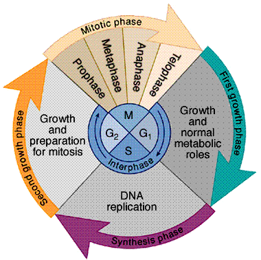
STAGE
1: Interphase
Much of a cell's
lifetime is spent undergoing cell growth, making proteins, sugars, and fats,
and assembling genetic material. This time in a cell cycle is called
interphase. Interphase consists
of three phases: G1, S, and G2.
|
PHASES
OF INTERPHASE |
||
|
G1 |
First
Gap Phase |
This is the phase when
the cell grows or increases in mass. The cell is producing proteins from the instructions
found in DNA and it is functioning as it is meant to. Cells that will not
divide stay in this phase (sometimes called G0 phase) and do not
move on. |
|
S |
Synthesis
Phase |
Synthesis refers to
making or producing. This is the phase when DNA is copied. The DNA is still
in the form of chromatin, but remember that this DNA with its copy attached
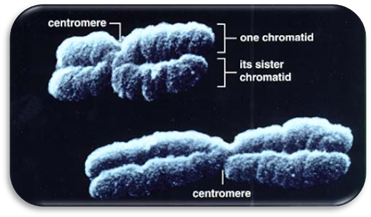
will make up a chromosome consisting of two chromatids held together by a
centromere as it continues to condense. |
|
G2 |
Second
Gap Phase |
This is the phase when
the cell continues to grow and prepares for cell division by producing
organelles, molecules, and structures called centrioles (which will be
discussed in a moment) to help with cell division. |
|
Visit Cells alive! To
see an animation of the cell cycle. http://www.cellsalive.com/cell_cycle.htm |
||
STAGE 2: Mitosis
|
STAGES OF MITOSIS |
|||
|
Order |
Stage |
Events |
Figure of Stage (onion root tip) |
|
#1 |
Prophase |
· Chromosomes begin
to condense and become visible. · The mitotic
spindle forms. · Centrioles move
to opposite poles. · Nuclear envelope
dissolves. |
|
|
In
prophase, the chromosomes begin to condense and become visible under a
microscope. Remember the DNA made a copy of itself in the S phase of
interphase and had the copy attach to the original by the centromere, but it
still needed to condense more. The mitotic spindle forms. The spindle is
responsible for moving the chromosomes around the inside of the cell. The
spindle, when fully formed, is described as being football-shaped when considering
a three-dimensional cell. Centrioles,
structures made of microtubules, will anchor the mitotic spindle. The
centrioles are constructed in G2 of interphase. Lastly, in
prophase the nuclear envelope dissolves. So, for a short time there is no nucleus. |
|||
|
#2 |
Metaphase |
· Centromeres line
up on the equator of the cell. |
|
|
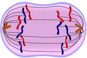
In
metaphase, the centromeres line up on the equator of the cell. The equator is the middle of the cell.
Sometimes the equator is called the metaphase
plate. The kinetochore fibers are responsible for dragging the
centromeres, which hold the two sister chromatids together, to the equator. |
|||
|
#3 |
Anaphase |
· Centromeres
separate pulling daughter chromosomes to opposite poles. |
|
|
In
anaphase, the kinetochore fibers split the centromere. By splitting the
centromere the two sister chromatids are now separated. The moment the sister
chromatids are separated in anaphase their name changes to daughter chromosomes. It is just a
name change. The daughter chromosomes are still identical copies of DNA, just
not connected by the centromere any more. The daughter chromosomes tend to
take on a V-shape because they are being dragged to opposite poles of the
cell through the cytoplasm. Sort of like dragging a straight piece of thread
through jelly from the middle of the thread. |
|||
|
#4 |
Telophase |
· Chromosomes
transition back to chromatin. · The mitotic
spindle dissolves. · Centrioles
dissolve · Nuclear envelope
reappears. |
|
|
Telophase
is practically the opposite of prophase. The daughter chromosomes uncoil to
become chromatin again. The mitotic spindle dissolves and will not be visible
again until the next mitotic division. Along with the spindle, the centrioles
are gone. The nuclear envelope reappears, therefore the nucleus is now
present and will be until the next mitotic division. |
|||
|
|
|||
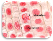
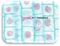
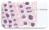
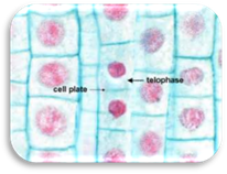
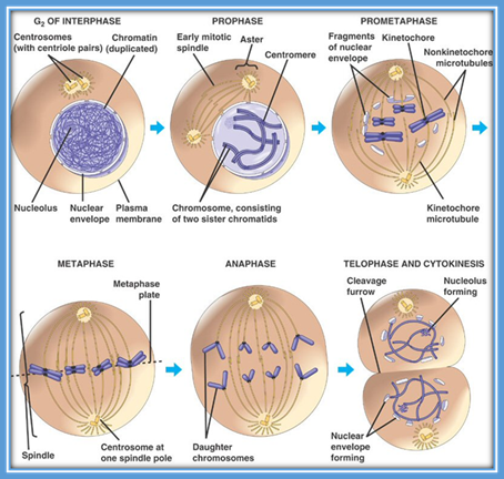
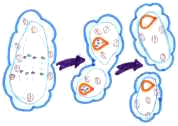
Figure of Mitosis in an Animal Cell
STAGE 3: Cytokinesis
 Mitosis
and its four stages mostly involved the DNA of the cell splitting into two new nuclei.
So the best way to define mitosis, which was mentioned in the beginning of this
unit, is to say mitosis is nuclear
division or division of the nucleus. Cytokinesis is concerned with
splitting everything else in the cell. Remember that everything else outside
the nucleus is cytoplasm, so, cytokinesis can best be defined as cytoplasmic division or division of the
cytoplasm.
Mitosis
and its four stages mostly involved the DNA of the cell splitting into two new nuclei.
So the best way to define mitosis, which was mentioned in the beginning of this
unit, is to say mitosis is nuclear
division or division of the nucleus. Cytokinesis is concerned with
splitting everything else in the cell. Remember that everything else outside
the nucleus is cytoplasm, so, cytokinesis can best be defined as cytoplasmic division or division of the
cytoplasm.
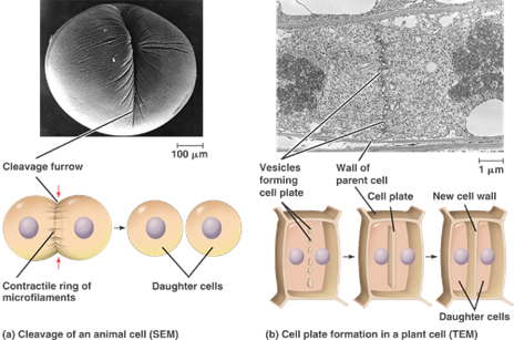
The process of
cytokinesis overlaps the end of mitosis. While mitosis finishes dividing the nucleus,
cytokinesis begins separating the cytoplasm. When cytokinesis begins a cleavage
furrow becomes present. A cleavage
furrow is the indentation in the middle of the cell membrane which is
attempting to pinch the cell in half. Animal cells and other eukaryotes without
a cell wall conduct cytokinesis in this fashion. Eukaryotes with a cell wall
cannot form a cleavage furrow so instead they have vesicles which will deposit
materials across the equator of the cell and form a new cell wall and cell
membrane. This new forming cell wall is called a cell plate. When cytokinesis finishes two new daughter cells have
formed and they will each enter their own G1 of interphase. It is
very important to remember that each daughter cell is identical to each other
and to the parent cell they came from.
![]() The Cell Cycle and Mitosis
The Cell Cycle and Mitosis
CANCER
 Cancer is essentially uncontrolled cell division.
It is a situation where cells are growing uncontrollably and sometimes
spreading. A growth of cancer cells is known as a tumor. A tumor is a growth caused by cells with an abnormal rate of
cell division and structure, and cells that lack a function. Cancer is mainly
due to some change in the DNA that controls the cell cycle making the cycle now
uncontrollable. There are two types of growth, or tumor, possible with cancer
cells. A benign tumor does not
spread to other parts of the body. Benign tumors can often be surgically
removed from the body. Malignant tumors
spread and destroy nearby healthy tissues and organs. The spreading of these
cancer cells is called metastasis.
Once cancer cells begin to spread it is very difficult to treat. Some tumors can
be treated by removing the affected organ, others require treatments to try to
control the cells. Many treatments, such as chemotherapy, aim to kill the
fast-growing cancer cells which also unfortunately affects healthy cells in the
process. Click on the image on the left to view an animation of cancer cells.
Cancer is essentially uncontrolled cell division.
It is a situation where cells are growing uncontrollably and sometimes
spreading. A growth of cancer cells is known as a tumor. A tumor is a growth caused by cells with an abnormal rate of
cell division and structure, and cells that lack a function. Cancer is mainly
due to some change in the DNA that controls the cell cycle making the cycle now
uncontrollable. There are two types of growth, or tumor, possible with cancer
cells. A benign tumor does not
spread to other parts of the body. Benign tumors can often be surgically
removed from the body. Malignant tumors
spread and destroy nearby healthy tissues and organs. The spreading of these
cancer cells is called metastasis.
Once cancer cells begin to spread it is very difficult to treat. Some tumors can
be treated by removing the affected organ, others require treatments to try to
control the cells. Many treatments, such as chemotherapy, aim to kill the
fast-growing cancer cells which also unfortunately affects healthy cells in the
process. Click on the image on the left to view an animation of cancer cells.
UNIT VOCABULARY
REVIEW
Click on the Quizlet link below to access the quizlet.com vocabulary flash cards.
Review the vocabulary before completing your assessment.

 Now answer questions 1 through 25.